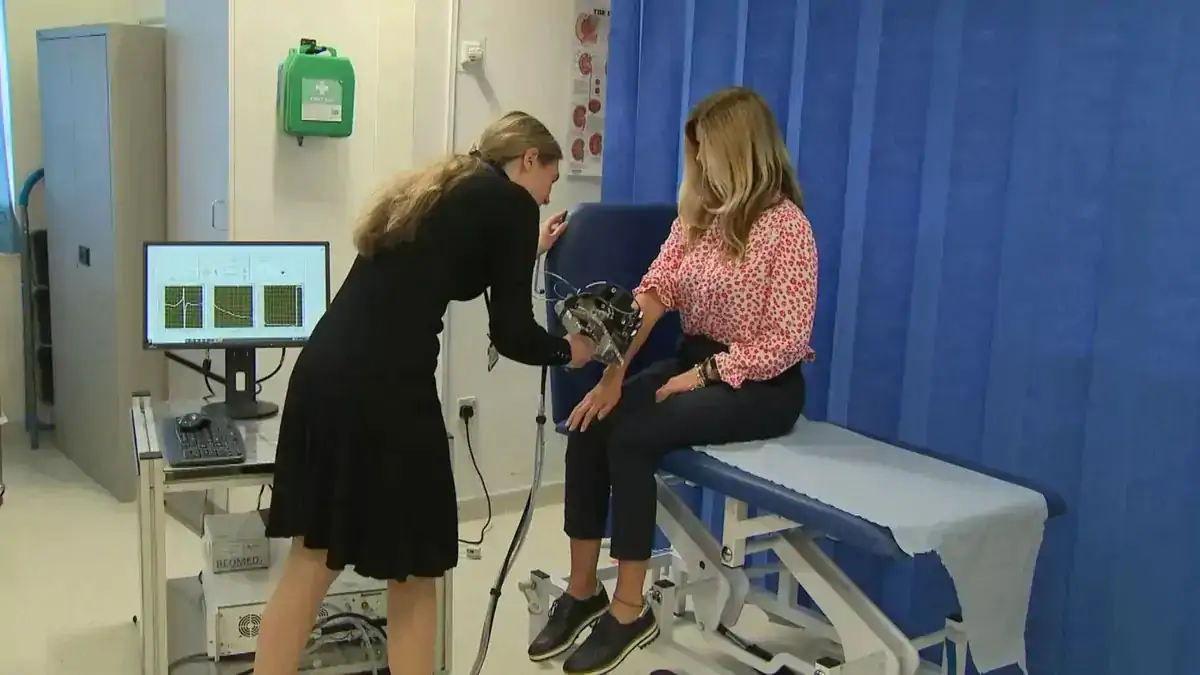
Professor Emma MacPherson is developing a new screening device that could revolutionize the diagnosis and treatment of skin cancers.
Skin cancer cases around the world
Skin cancer is one of the most common and deadly forms of cancer, affecting millions of people worldwide. Early detection and treatment are crucial to improving patients' survival rates and quality of life. However, current methods of diagnosis, such as biopsies and dermatoscopes, are invasive, expensive, and sometimes inaccurate.
That is why Professor Emma MacPherson, a physicist at the University of Warwick, is working on a novel technology that could change how skin cancers are detected and screened. She uses terahertz radiation, a type of electromagnetic wave between microwaves and infrared light, to create images of the skin that reveal changes in hydration and blood flow.
How radiation has several advantages
Terahertz radiation has several advantages over other imaging modalities, such as X-rays and ultrasound. It is non-ionizing, meaning it does not damage the cells or tissues of the body. It is also sensitive to water, which makes it ideal for detecting skin cancers, as they tend to have different water content and blood supply than normal skin.
Professor MacPherson and her team at the Department of Physics are developing a screening device that uses terahertz frequencies to scan the skin and produce high-resolution images that can identify suspicious lesions. The device is portable, fast, and easy to use and could be deployed in clinics, hospitals, and pharmacies.

Professor MacPherson said, "We have been working on this technology for over 20 years, and we are now at the stage where we can test it on real patients in the clinical setting. Our goal is to provide a non-invasive, low-cost, and accurate tool that can help diagnose skin cancers at an early stage and guide the best treatment options."
Professor MacPherson's achievements
Professor MacPherson is not only a pioneer in the field of terahertz imaging but also a role model for women and girls in science. She recently became the first-ever female and youngest recipient of the International Society of Infrared, Millimeter, and Terahertz Waves (IRMMW-THz) Society's Exceptional Service Award, recognizing her outstanding contributions to the scientific community.
She has organized and hosted several international conferences and workshops on terahertz research and has supported young researchers, especially women, in the field. She is also one of the founders of the Zhenyi Wang Award, which honors young women who excel in terahertz research.
Professor MacPherson said, "I am delighted and honored to receive the Exceptional Service Award from the IRMMW-THz Society. I have always felt welcomed and encouraged by the terahertz community, and I hope to inspire and mentor the next generation of scientists, especially women and girls, who want to pursue a career in this exciting and emerging field."
Professor MacPherson's latest research paper on terahertz imaging of skin cancers is published this week on the Advanced Photonics Nexus, ahead of the International Day of Women and Girls in Science on February 11.
Study abstract:
This study introduces a handheld terahertz (THz) scanner designed to quantitatively evaluate human skin hydration levels and thickness. This device, through the incorporation of force sensors, demonstrates enhanced repeatability and accuracy over traditional fixed THz systems. The scanner was evaluated in the largest THz skin study to date, assessing 314 volunteers, successfully differentiating between individuals with dry skin and hydrated skin using a numerical stratified skin model. The scanner measures and displays skin hydration dynamics within a quarter of a second, indicating its potential for real-time, noninvasive examinations, opening up opportunities for in vivo and ex vivo diagnosis during patient consultations. Furthermore, the portability and ease of use of our scanner enable its widespread application for in vivo and ex vivo diagnosis during patient consultations, potentially allowing in situ biopsy evaluation and elimination of histopathology processing wait times, thereby improving patient outcomes by facilitating simultaneous tumor diagnosis and removal.


 BlocksInform


